Introduction: Ever Wondered How Doctors Read an ECG?
Have you ever looked at an ECG (electrocardiogram) and thought, “What on earth is that chart saying about my heart?” If you’re curious about how doctors interpret these squiggly lines, you’re in the right place.
ECGs are crucial for diagnosing heart conditions, but reading them can seem overwhelming at first. Don’t worry! In this guide, we’ll break down how to read an ECG step by step, so you can start recognizing key patterns and understand what your heart is telling you.
What is an ECG?
How to Read an ECG: Understanding the Basics of an ECG
An ECG (or EKG) is a test that measures the electrical activity of your heart over a period of time. It provides valuable insights into how well your heart is working and helps doctors diagnose a range of heart conditions, from arrhythmias to heart attacks.
Imagine your heart as an electrical system. Every beat is controlled by electrical impulses that travel through the heart, causing it to contract and pump blood. The ECG captures these electrical signals as waves on a graph, which doctors can analyze.
How Does an ECG Work?
ECGs are done by placing small electrodes on your skin, usually on your chest, arms, and legs. These electrodes detect the electrical signals from your heart and send them to a machine that creates the ECG waveform.
The Key Components of an ECG
1. Heart Rate
Before diving into the waveforms, one of the first things to check on an ECG is the heart rate. You can calculate it by counting the number of R waves (the sharp upward spikes) in a 6-second strip and multiplying by 10.
- Normal heart rate: 60-100 beats per minute (bpm)
- Tachycardia: Heart rate over 100 bpm
- Bradycardia: Heart rate under 60 bpm
2. Rhythm
Is the rhythm regular or irregular? This is a crucial first step in ECG interpretation.
- Regular rhythm: The time between beats is consistent.
- Irregular rhythm: The time between beats varies, which can signal arrhythmias.
3. P Wave
The P wave represents atrial depolarization, which is the electrical impulse moving through the atria (upper chambers of the heart).
- A normal P wave is small and rounded, indicating normal atrial activity.
- Abnormal P waves can signal problems like atrial fibrillation, where the rhythm is irregular and there may be no clear P waves.
4. QRS Complex
The QRS complex represents ventricular depolarization, or the electrical impulse traveling through the ventricles (lower chambers of the heart). This is the most important part to focus on when assessing the ECG’s overall rhythm and function.
- Normal QRS: Narrow, sharp, and less than 120 milliseconds in duration.
- Wide QRS: A duration longer than 120 ms, which could indicate a bundle branch block or other conduction issues.
5. T Wave
The T wave represents ventricular repolarization—the process by which the ventricles “reset” after contracting.
- Normal T waves are upright and symmetrical.
- Abnormal T waves can indicate issues like ischemia (lack of blood flow), electrolyte imbalances, or myocardial infarction (heart attack).
How to Interpret an ECG: A Step-by-Step Guide
Step 1: Check the Heart Rate
Start by measuring the heart rate. This can help you quickly determine if the person is experiencing tachycardia (fast heart rate) or bradycardia (slow heart rate). Simply count the R waves in a 6-second strip and multiply by 10 for an estimate of the heart rate.
Step 2: Look at the Rhythm
Next, assess whether the rhythm is regular or irregular. Is the distance between R waves equal, or do they vary?
- Regular rhythm: This is typically normal.
- Irregular rhythm: This might indicate an arrhythmia, such as atrial fibrillation.
Step 3: Examine the P Waves
The P wave should be positive (upright) in lead II, indicating that the atria are contracting properly.
- Absent P waves: This could point to atrial fibrillation or other abnormal rhythms.
- Abnormal P waves: In cases like atrial enlargement, you might see a peaked or bifid P wave.
Step 4: Analyze the QRS Complex
The QRS complex should be sharp and narrow. A wide QRS (greater than 120 ms) suggests delayed conduction through the ventricles, which could be caused by a bundle branch block or other conduction problems.
- Normal QRS: Narrow and sharp.
- Wide QRS: Could indicate ventricular tachycardia or conduction delays.
Step 5: Check the T Waves
Examine the T waves for abnormal shapes or inverted waves. An inverted T wave can be a sign of ischemia or myocardial infarction. Tall, peaked T waves might indicate an electrolyte imbalance, particularly hyperkalemia (high potassium levels).
Common ECG Patterns and Their Meanings
1. Sinus Rhythm: A Healthy Heart
In a normal ECG, the heart rhythm follows a regular, consistent pattern, and each beat begins in the sinus node, the heart’s natural pacemaker.
- Sinus rhythm: This means the heart is beating in a regular, healthy way.
2. Atrial Fibrillation (AFib)
One of the most common arrhythmias, atrial fibrillation causes an irregular, often rapid heart rate. On an ECG, you will notice:
- Irregular rhythm
- Absent P waves
- Rapid ventricular response (RVR)
AFib can lead to strokes, so it’s important to catch it early.
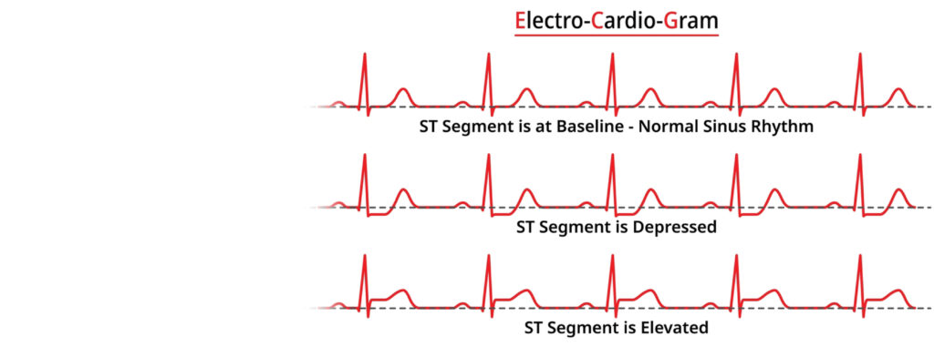
3. Myocardial Infarction (Heart Attack)
One of the most important things to look for on an ECG is ST elevation or depression, which can indicate a heart attack. A significant ST elevation in specific leads (like leads II, III, and aVF) often signals a STEMI (ST-elevation myocardial infarction), a type of heart attack requiring immediate treatment.
4. Ventricular Tachycardia (VT)
Ventricular tachycardia is a potentially life-threatening arrhythmia where the heart beats too fast from the ventricles. On an ECG, you’ll see:
- Wide QRS complexes
- Rapid, regular rhythm
VT requires urgent medical attention.
5. Long QT Syndrome
Long QT syndrome is a condition where the QT interval (the time between the Q wave and the T wave) is prolonged. This can lead to dangerous arrhythmias, such as torsades de pointes.
Tips for Beginners
Practice with Real ECGs
The more you practice reading ECGs, the better you’ll get. Look for real ECG strips online or in textbooks, and try to identify rhythms and abnormalities on your own.
Use a Systematic Approach
ECG interpretation can be overwhelming, but using a systematic approach makes it easier. Start with the heart rate, then check the rhythm, followed by the P wave, QRS complex, and T wave.
Don’t Rush—Take Your Time
Reading an ECG is a skill that takes time to develop. Don’t be discouraged if you don’t catch every detail right away. With patience and practice, you’ll improve.
Conclusion: Ready to Start Interpreting ECGs?
ECGs are a powerful tool for understanding heart health, and with a bit of practice, you can start interpreting these patterns yourself. Whether you’re learning for professional reasons or just curious about how your heart works, breaking down the ECG into manageable parts makes it much easier to understand.
According to Wikipedia, an electrocardiogram (ECG or EKG) is a medical test that measures the electrical activity of the heart over time. It records the timing of electrical signals as they pass through the heart, helping doctors assess heart function, diagnose heart conditions, and monitor heart health. The test is performed by placing electrodes on the skin, which detect the electrical impulses produced by heartbeats. ECGs are commonly used to detect irregularities in heart rhythm, signs of heart attacks, and other cardiovascular problems. For a more detailed explanation, you can visit the Wikipedia page on Electrocardiography.
FAQ: Common Questions About Reading ECGs
1. Can I interpret an ECG without medical training?
While it’s possible to learn the basics of ECG interpretation, a full understanding requires medical training. Start with the basics and practice regularly to improve.
2. What should I do if I notice something unusual on my ECG?
If you notice something unusual, it’s always a good idea to consult a doctor or a trained healthcare professional to confirm the findings.
3. Can an ECG detect all heart problems?
An ECG can detect many heart conditions, but not all. It’s a valuable tool, but some conditions may require other diagnostic tests, like an echocardiogram or stress test.
What’s Your Experience with Reading ECGs?
Now that you have the basics, what’s your experience with interpreting ECGs? Have you seen any common patterns or tricky moments? Let us know in the comments below!
