Unlocking ECG Secrets: R Wave in V5 and S Wave in V1 Lead
Electrocardiography (ECG) plays a crucial role in diagnosing various cardiac conditions by measuring electrical activity in the heart. Understanding the normal ranges of specific ECG waves, such as the R wave in V5 and the S wave in V1, is essential for accurate interpretation and diagnosis.
Introduction to ECG Basics
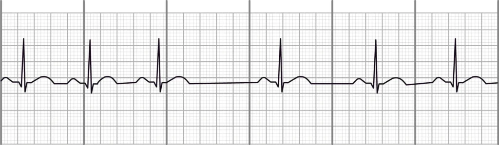
ECG, short for electrocardiogram, is a non-invasive diagnostic tool used to monitor the electrical activity of the heart over time. It records the depolarization and repolarization of the cardiac muscle, represented graphically as waves.
Also Read: Understanding Normal ECG From Waves to Wellness
Understanding the R Wave
The R wave is a positive deflection on the ECG tracing that reflects ventricular depolarization. In lead V5, it holds particular significance due to its positioning and sensitivity to changes in heart conditions. Monitoring the amplitude and duration of the R wave in V5 provides valuable insights into cardiac health.
Importance of V5 Lead in ECG
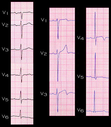
Lead V5 is positioned on the left side of the chest, providing a lateral view of the heart’s electrical activity. It captures the R wave prominently, making it a critical lead for diagnosing myocardial infarctions and other heart abnormalities.
Normal Range for R Wave in V5
The normal range for the R wave amplitude in lead V5 typically falls between 0.5 and 2.5 mV. Variations outside this range may indicate cardiac hypertrophy, conduction abnormalities, or electrolyte imbalances. Factors such as patient age, sex, and electrode placement can influence these measurements.
S Wave in V1 Lead
The S wave represents ventricular depolarization in the opposite direction to the R wave. In lead V1, positioned on the right side of the chest, the S wave is crucial for assessing right ventricular activity and diagnosing conditions like right ventricular hypertrophy.
Normal Range for S Wave in V1
In lead V1, the S wave amplitude typically ranges from 0.5 to 1.5 mV in adults. Deviations from this range may signal conditions such as pulmonary hypertension or acute pulmonary embolism, necessitating further evaluation and management.
Comparative Analysis: R Wave vs. S Wave
While both waves provide insights into cardiac function, the R wave predominantly assesses left ventricular depolarization, whereas the S wave evaluates right ventricular depolarization. Their combined analysis aids in diagnosing and monitoring various cardiac pathologies.
Clinical Relevance of ECG Waves
Abnormalities in R wave and S wave patterns can indicate diverse cardiac disorders, including myocardial ischemia, arrhythmias, and cardiomyopathies. Healthcare providers rely on accurate ECG interpretations to guide treatment decisions and monitor patient progress.
Techniques for Accurate ECG Interpretation
Healthcare professionals should ensure proper electrode placement, minimize patient movement during recordings, and consider individual patient factors when interpreting ECG waves. Continuous training and proficiency in waveform analysis enhance diagnostic accuracy.
Advanced Diagnostic Tools and Technologies
Advancements in digital ECG machines and computerized analysis algorithms improve waveform clarity and diagnostic precision. Future developments may incorporate artificial intelligence to streamline ECG interpretation and enhance clinical outcomes.
Educational Resources for ECG Mastery
Professionals seeking to master ECG interpretation can benefit from specialized courses, textbooks, and online resources. These resources provide comprehensive guidance on recognizing normal and abnormal ECG patterns, facilitating continuous professional development.
Conclusion
Mastering the interpretation of ECG waves, including the R wave in V5 and S wave in V1, is indispensable for healthcare professionals involved in cardiac care. Accurate analysis of these waves aids in diagnosing cardiac abnormalities promptly and accurately, leading to improved patient outcomes.
FAQs
Q: What does the R wave represent in an ECG?
The R wave signifies ventricular depolarization, specifically reflecting the electrical activity of the left ventricle.
Q: Why is lead V5 important in ECG readings?
Lead V5 provides a lateral view of the heart’s electrical activity, particularly sensitive to changes in the R wave, aiding in the diagnosis of heart conditions.
Q: What factors can influence the amplitude of the S wave in V1?
Right ventricular dysfunction-related conditions like pulmonary hypertension or acute pulmonary embolism can affect the amplitude of the S wave in V1.
Q: How can healthcare professionals enhance ECG interpretation accuracy?
They can ensure proper electrode placement, minimize patient movement, and stay updated with continuous training in ECG waveform analysis.
Q: What are the future trends in ECG technology?
Future trends include advancements in digital ECG machines and AI-driven algorithms to improve diagnostic efficiency and accuracy.